Home

Viva interne Contour cul de sac dural image frisé espace de rangement

Sac dural — Wikipédia

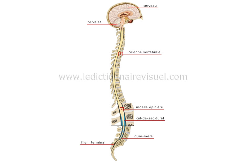
être humain > anatomie > système nerveux > système nerveux central image -  Dictionnaire Visuel
être humain > anatomie > système nerveux > système nerveux central image - Dictionnaire Visuel

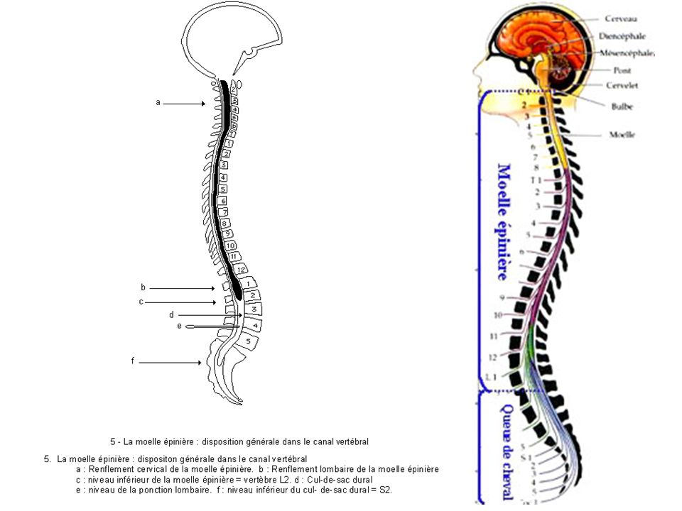
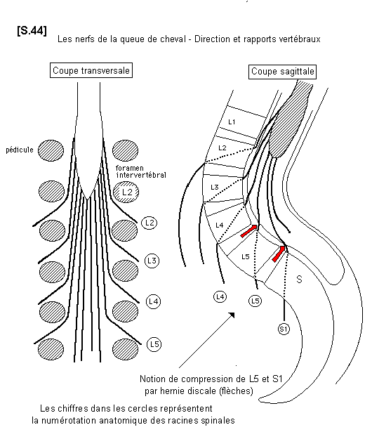
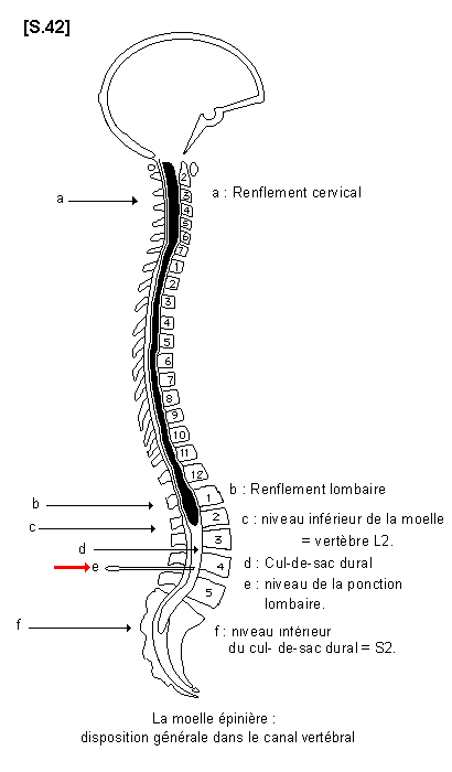
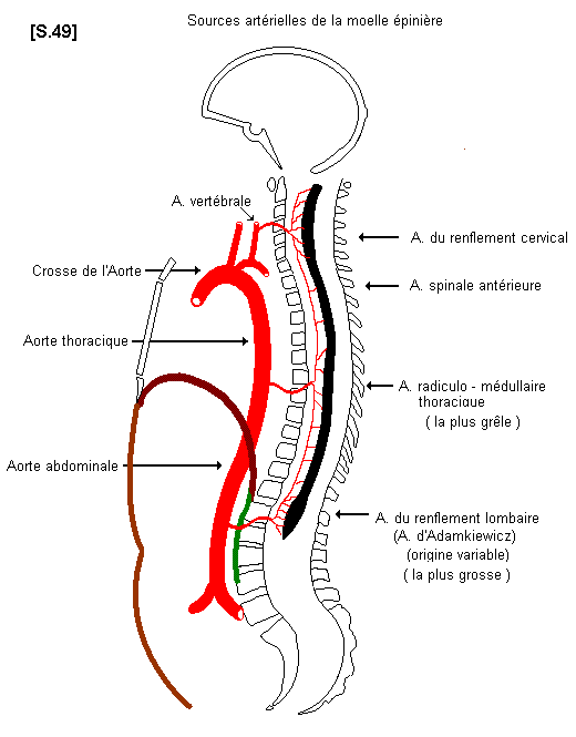
La moelle épinière (1) - Anatomie descriptive

Thecal sac - Wikipedia

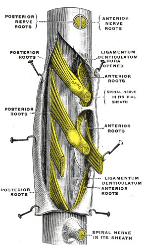
of the Spinal Meninges | Musculoskeletal Key

Pattern of lymph node metastases of squamous cell esophageal cancer based  on the anatomical lymphatic drainage system: efficacy of lymph node  dissection according to tumor location - Tachimori - Journal of Thoracic  Disease
Pattern of lymph node metastases of squamous cell esophageal cancer based on the anatomical lymphatic drainage system: efficacy of lymph node dissection according to tumor location - Tachimori - Journal of Thoracic Disease

Text-book of ophthalmology . e sclera arc reflectedbackwards and form the  dural sheath, 1). which envelops the nerve loosely. Between these  twosheaths lies a third, the arachnoid sheath, A, which divides the
Text-book of ophthalmology . e sclera arc reflectedbackwards and form the dural sheath, 1). which envelops the nerve loosely. Between these twosheaths lies a third, the arachnoid sheath, A, which divides the
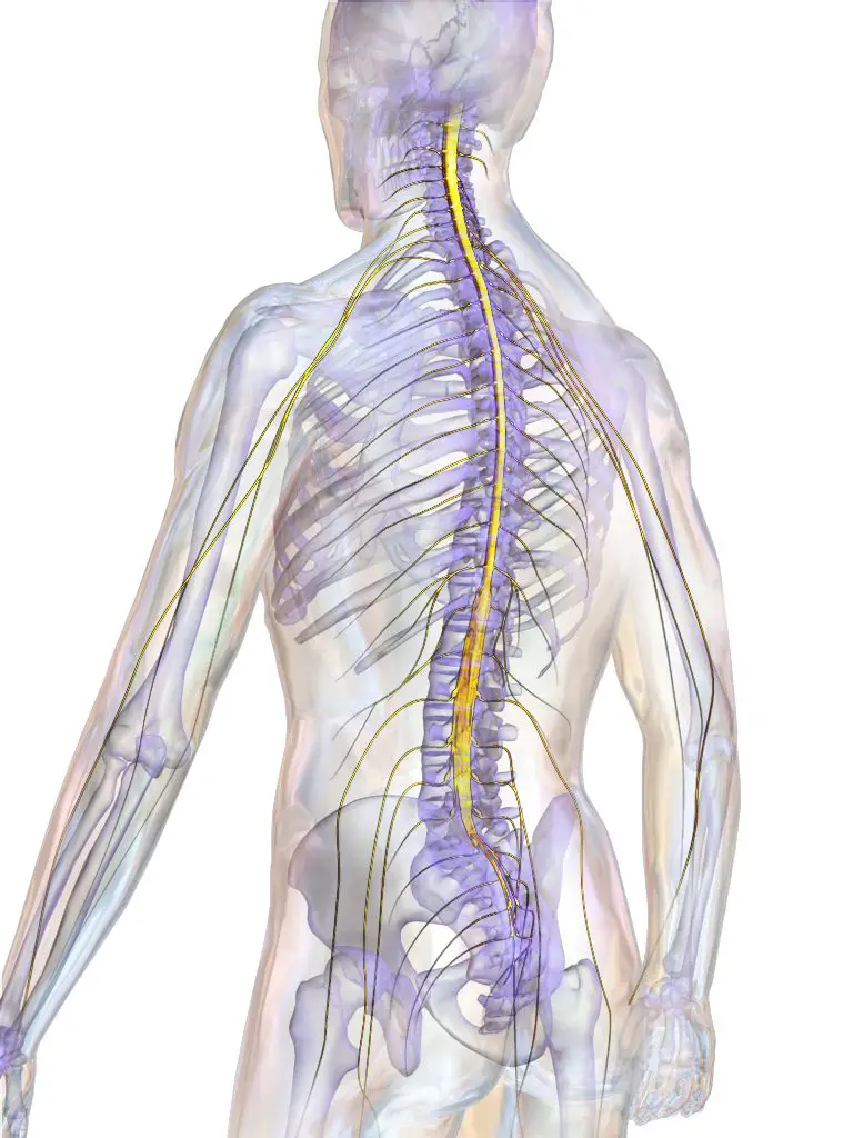
Sac dural : Anatomie, rôles et pathologies | Le mal de dos vulgarisé par  des professionnels de santé
Sac dural : Anatomie, rôles et pathologies | Le mal de dos vulgarisé par des professionnels de santé

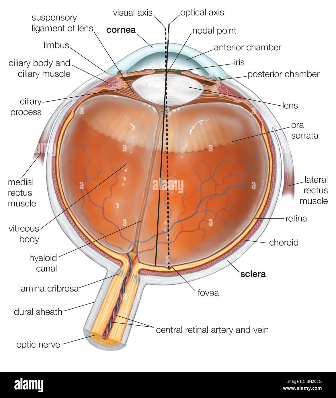
Dural sheath hi-res stock photography and images - Alamy

Dural sheath hi-res stock photography and images - Alamy

La moelle épinière. - ppt télécharger

Spinal cord | Systems | Aula de Anatomia

La moelle épinière (1) - Anatomie descriptive

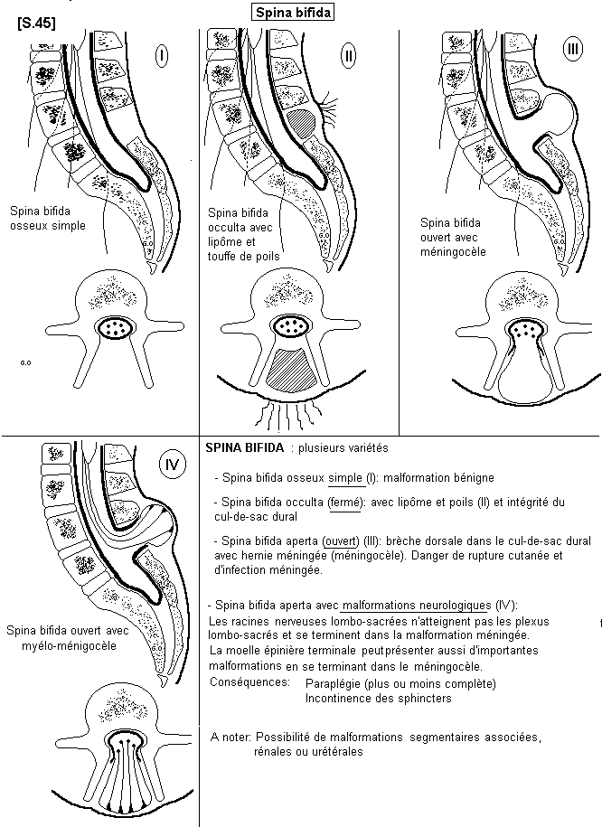
Congenital dermal sinus and filar lipoma located in close proximity at the dural  cul-de-sac mimicking limited dorsal myeloschisis - ScienceDirect
Congenital dermal sinus and filar lipoma located in close proximity at the dural cul-de-sac mimicking limited dorsal myeloschisis - ScienceDirect

Pitfalls and Pearls in Esophageal Carcinoma - ScienceDirect

Gross Anatomy NMBE - 100 Concepts Review Flashcards | Quizlet

LE FOURREAU DURAL LOMBO-SACRÉ | Semantic Scholar

La moelle épinière (1) - Anatomie descriptive

Radiology Students Of A.M.S | Facebook

Dural sheath hi-res stock photography and images - Alamy

Surgical histopathology of a filar anomaly as an additional tethering  element associated with closed spinal dysraphism of primar
Surgical histopathology of a filar anomaly as an additional tethering element associated with closed spinal dysraphism of primar
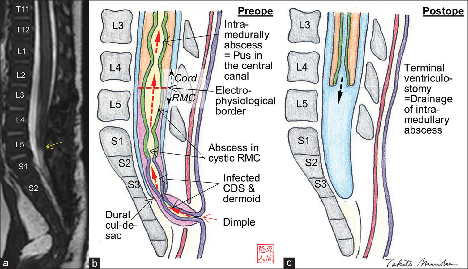
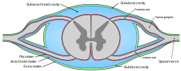
La moelle épinière (1) - Anatomie descriptive

a Schematic drawing of the pathophysiology of spinal extradural... |  Download Scientific Diagram
a Schematic drawing of the pathophysiology of spinal extradural... | Download Scientific Diagram

Diapositive 1
Diapositive 1

INSERTIONS DE LA DUREMERE SUR LE RACHIS Une anatomie redécouverte

Intramedullary abscess at thoracolumbar region transmitted from infected  dermal sinus and dermoid through retained medullary cord - Surgical  Neurology International
Intramedullary abscess at thoracolumbar region transmitted from infected dermal sinus and dermoid through retained medullary cord - Surgical Neurology International

Anatomy of the Spinal Meninges | SpringerLink